APEXVIEW APX100 Benchtop Fluorescence Microscope
The APEXVIEW™ APX100 benchtop fluorescence microscope delivers the imaging capabilities researchers need to generate results that advance research. Designed around the realities of modern laboratories, it combines simplicity, performance, image quality, and flexibility in a fully digital platform. Intelligent automation reduces manual, error-prone tasks, while premium optics and powerful software help provide reliable data researchers can trust.
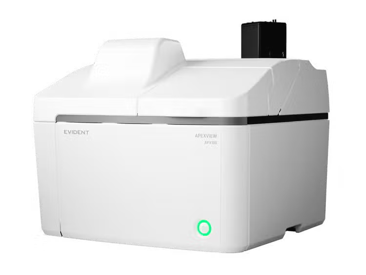
APX100 Benchtop Fluorescence Microscope
Results That Advance Research
Researchers need to focus on discovery, not equipment.
The APX100 benchtop fluorescence microscope automates imaging, from finding samples and autofocusing to managing data. With a user-friendly interface, every capability is accessible in just a few clicks, with no separate expert mode required.
The fully digital system eliminates the complexity of conventional microscopy controls and the need for a darkroom, making advanced imaging accessible to researchers of all experience levels.
Capture samples with multichannel fluorescence, brightfield, image stitching, Z-stack imaging, time-lapse imaging, and gradient contrast—all on a single platform that moves discovery forward.

Mouse kidney. Alexa Fluor 488 WGA, Alexa Fluor 568 Phalloidin, DAPI.
Intelligent Automation
Simplicity That Empowers Users
Automatic Sample Detection
The APX100’s smart sample navigator uses built-in AI to automatically locate samples and regions of interest, helping you get to imaging and data collection faster. Just select your sample, choose how you want to image, and start capturing data.
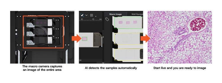
High-Speed Autofocus
The APX100’s autofocus is up to 12 times faster than conventional autofocus algorithms, so you can spend less time adjusting and more time collecting data.
Intuitive Interface
Using the APX100 is simple—load the sample, close the lid, and click a button. The intuitive software simplifies imaging workflows with an easy-to-learn interface, enabling you to set up complex multidimensional experiments with confidence.

Automated Project and Data Management
The APX100 automatically labels, organizes, and stores images, helping reduce manual effort and minimize the risk of errors.
- Organized, easy-to-find images: During image acquisition, the software automatically creates folders for each sample and saves the data into the correct one
- Consistent indexing: Keeps your data organized and easy to locate, preventing accidental saving of data in the wrong folder
- Recall image acquisition settings: All important acquisition settings are saved alongside image data so that they can be easily recalled for future experiments
Insightful Images
Images That Drive New Insights
Scientific breakthroughs depend on accurate, quantifiable data.
The APX100 combines premium optical hardware with powerful image processing and analysis, giving your research the high-quality images and enabling the deeper insights your work demands.
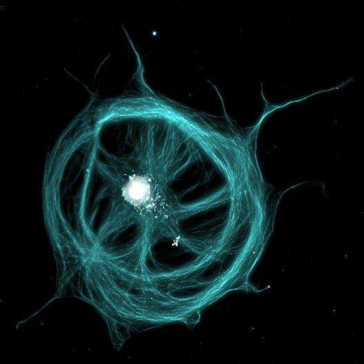
Tubulin and nucleus of BSC-1 cells.


Rat brain slice. Left: Spherical aberration is causing blur. Right: Spherical aberration corrected with the motorized correction collar.
Optics Tailored to Your Research
Evident’s renowned optics help ensure you see true sample details, supporting an accurate understanding of the location and makeup of biological structures.
Equip the APX100 with our award-winning X Line™ objective series for bright, consistent imaging, or use our application-specific objectives designed to meet any research need. Advance your discoveries with options for silicone immersion, long working distance, and plastic correction.
A motorized correction collar can tune the objective lens to your sample type for further improved image quality.
Clearly View Unstained Samples
The APX100’s gradient contrast method provides high-contrast, detailed visualization of unstained specimens.
- Clear visualization without staining: Observe anything from thick samples to small cellular structures
- Reduces the risk of contamination: Image through both glass and plastic dishes and multi-well plates
- Consistent clarity: Reduces halo effects and other visual artifacts that can occur with conventional phase visualization methods
"Gradient Contrast significantly aided in verifying disease reproducibility by clearly capturing the fine structure of organoids. Furthermore, its ability to enable high-quality observation under routine culture conditions is another compelling feature."
Toshiro Sato MD/PhD
Ryo Igarashi MD/PhD
Keio University
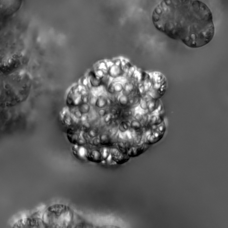
Gradient contrast imaging of hepatocyte organoids,
courtesy of Ryo Igarashi MD/PhD and Toshiro Sato MD/PhD, Keio University, School of Medicine
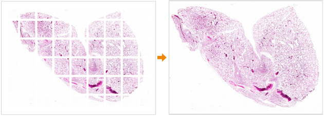
Mouse lung captured with a UPLXAPO4X objective. Stain: HE.
Seamlessly Stitched Images
See your whole sample in context. Evident's Intelligent Shading Correction automatically assembles large areas into one seamless image, with precise alignment and shading correction that keeps illumination consistent from edge to edge.
Powerful Image Processing
Evident's TruSight™ deconvolution recovers detail beyond the limits of the optics, sharpening and enhancing your images while preserving the quantitative integrity researchers rely on for confident analysis.


Left: Without TruSight. Right: With TruSight.
Slide courtesy of Encor Scientific.
Simplified Live-Cell Imaging
The APX100 supports long-term live-cell imaging with tools that sustain cell viability while delivering consistent image quality across every time point.
- Gradient contrast: Provides clear, detailed visualization of transparent specimens, thick samples, and cellular structures without staining
- Incubation chamber: Maintain stable culture conditions with controlled temperature, humidity, and CO₂
- Surface Finder: Combats focal drift to keep the sample in focus during long-term, time-lapse experiments
- Perfusion systems: Deliver compounds or exchange media mid-experiment to capture cellular response as it happens
Scratch assay
Scratch assay time-lapse imaging of Human Aoritic Enodothelial Cells (HAEC) (2 days).
Image data courtesy of Dr. Kazutaka Ueda, Department of Cardiovascular Medicine, The University of Tokyo Hospital.
Fertilized mouse egg
4-hours time-lapse observation of a fertilized mouse egg.
Image Processing and Analysis Functions that Accelerate Research
Our advanced cellSens image processing and analysis software works seamlessly with the APX100 system, providing you the tools to process and analyze your data quickly and efficiently.
cellSens Image Analysis
An optional cellSens software license provides powerful analysis capabilities.
- Access image processing techniques, including counting intracellular signals, analyzing the brightness of time-lapse data, and Evident's TruAI deep-learning technology
- The TruAI deep-learning solution offers flexibility, from predicting multi-class nuclei phenotypes for drug testing to rapid automated detection and segmentation of glomeruli
- cellSens integration enables easy, on-the-spot processing for simple measurements and image export
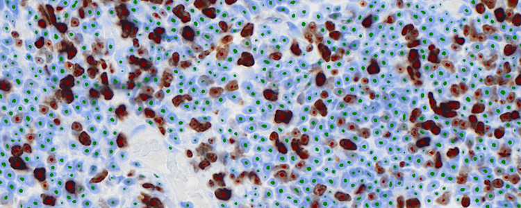
Cell & Colony Analysis
- Cell counting
- Colony counting
- Cell density estimation
- Area coverage / confluency estimation
- Size distribution analysis
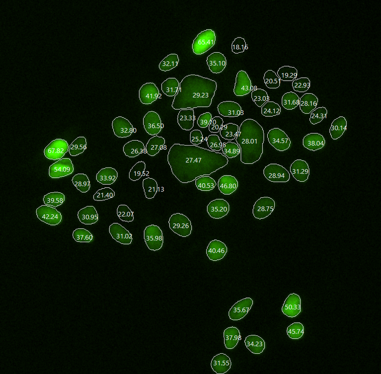
Morphology Analysis
- Cell morphology characterization
- Nuclear morphology analysis
- Circularity measurement
- Aspect ratio measurement
- Feret diameter analysis
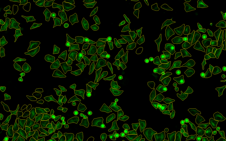
Fluorescence Quantification
- Reporter expression quantification
- Fluorescence intensity measurement
- Positive-cell analysis
- Multi-channel intensity comparison
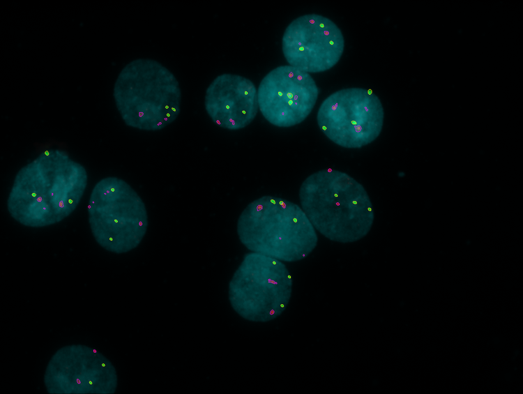
Spot & Puncta Analysis
- Fluorescent puncta counting
- Granule analysis
- Fluorescence spot analysis
- Aggregate quantification
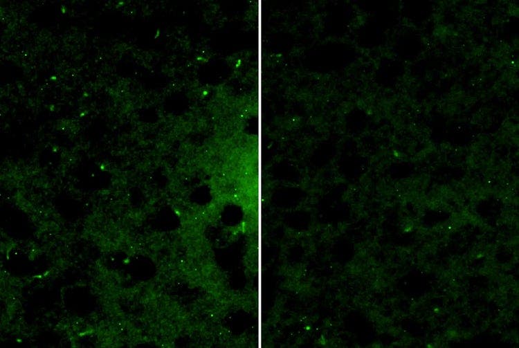
"We need a microscope capable of accurately imaging extremely fine details to capture structural changes in primary cilia. Among the observation tools that we have used, the APX100 microscope has performed exceptionally and reliably."
Dr. Yuki Kobayashi
Associate Professor, Hiroshima University
https://adobeassets.evidentscientific.com/content/dam/mis/apx100/videos/Fig2_movie_OriginalSize.mp4
Comparative images of primary cilia in the brains of control mice and mice subjected to 21 days of stress. Images courtesy of Professor Yuki Kobayashi, Hiroshima University.
Powerful Flexibility
"The APX100’s dual camera system—featuring both a high-sensitivity monochrome camera for fluorescence and a color camera for natural imaging—enables us to capture both types of images simultaneously. This capability is of great value in plant research."
Dr. Hirotsuna Yamada
Assistant Professor, Hiroshima University
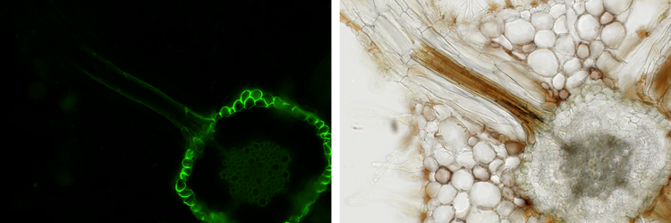
Fluorescence (left) and brightfield (right) images of mature cluster roots and non-cluster roots. Images courtesy of Dr. Hirotsuna Yamada.
Powerful for Everyone
Researchers need core-facility-level imaging without the barriers of access, or expertise. The APX100 combines automated scanning, intelligent screening, and advanced imaging tools in an easy-to-use platform that delivers powerful results for every user.
- Capture 4D images: Combine expanded field of view, depth, and time-lapse (or all three together) to get more from every sample than a single flat image can show
- Multiplex to the NIR: A broad wavelength range supports more simultaneous colors, better separation between markers, and visibility deeper into the sample
- Detect faint signals: The high-sensitivity camera's readout noise of just 0.7 electrons captures even dim biological signals as reliable data, without overexposing your sample
- Faster fluorescence imaging: A high-speed multi-LED light source captures multichannel images up to 2.4x faster*, so you can collect more data in less time
*Compared to a white LED light source. Tested collecting a 4-color image across a 10×10 multi-image acquisition.
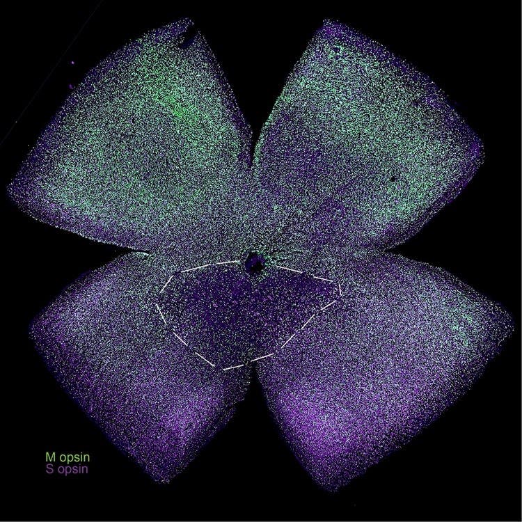
Mouse retina flat mount stained with anti S-opsin (violet) and M-opsin (green) antibodies. The dashed lines highlight an area of cone density deterioration due to light damage. Image courtesy of Dr. Henri Leinonen.
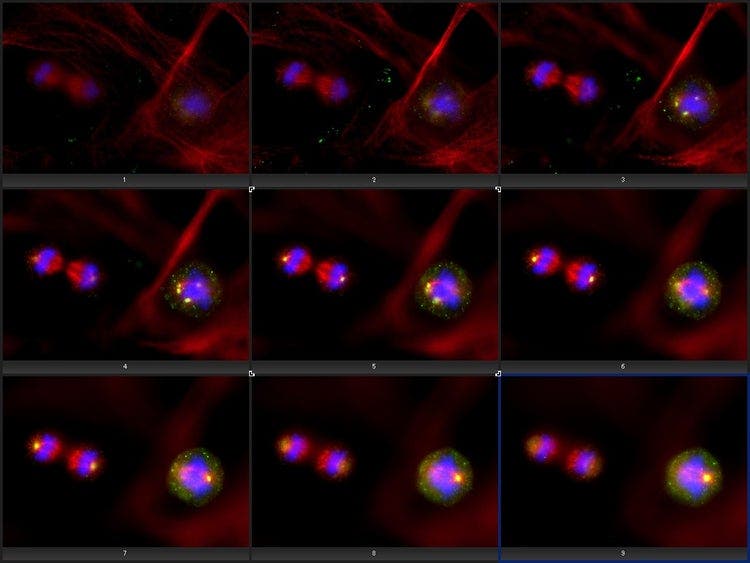
Subcellular localization of kendrin/pericentrin, centrosome protein. Image data courtesy of Kazuhiko Matsuo, PhD, Division of Developmental Biology and Anatomy, Department of Anatomy, Kyoto Prefectural University of Medicine.

“I have been genuinely pleased with the APX100 performance so far in a busy, diverse laboratory. It is feeding data to multiple in-house projects, as well as helping us set up capabilities for new academic collaborations. The options for upgrades are very good, with the advantage of a lower capital expenditure investment.”
Mark Taylor, PhD
Fermentation Lead, Marlow Ingredients
Fits Your Space, Your Samples, and What Comes Next
As research needs evolve, the APX100 adapts—protecting long-term investments and supporting scientific discovery for years to come.
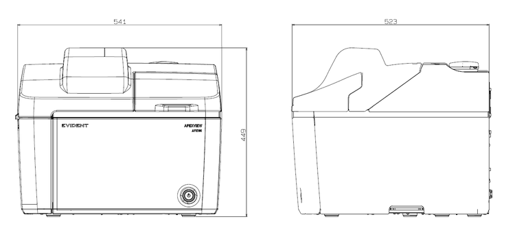
Research needs evolve, and lab space is always at a premium. The APX100 adapts with broad sample compatibility, a compact benchtop footprint, and upgradable capabilities, delivering the confidence to support today’s applications and the flexibility for tomorrow’s discoveries.
- Enclosed design: Fully sealed, so there's no need for a dedicated microscope room; it fits on any benchtop with a power outlet
- Built-in antivibration: Stable, high-resolution imaging on a standard lab bench—no separate antivibration table required
- Broad sample-holder compatibility: Supports most common formats labs use, from slides and well-plates to petri dishes
- Configurable light sources, cameras, filter cubes, and objectives: Customize the system for today's needs, and reconfigure it again as your requirements change
Specifications
| Category | Specification | APX100-HCU |
| Microscope | Observation method | Brightfield, fluorescence, phase contrast, gradient contrast |
| Sample holder | Glass slides (3 slides), 35 mm dish (3 dishes), microplate, general | |
| Objectives | Choose from 25 available objectives (4X–100X) | |
| Six-position motorized revolving nosepiece | ||
| Motorized aberration correction | One motorized position, five standard positions for objectives | |
| Transmitted illuminator | Built-in Köhler illumination for transmitted light, high color rendering LED | |
| Condenser: WD 45 mm, including PHL, PH1, PH2, and PH3 | ||
| Stage | Motorized XY stage with automatic control | |
| Focusing | Motorized focusing with automatic control | |
| Magnification changer | Color: 0.5x fixed; Monochrome: DP23M 0.5x fixed, Others 1x, 2x |
|
| Fluorescence | Fluorescence illuminator with fly-eye lens | |
| Eight-position motorized mirror turret | ||
| Evident U-LGPS (Class 1 laser product) | ||
| Motorized ND filter changer (100%, 25%, 6%) | ||
| Macro optical system | Built-in, 0.07x macro optics | |
| Anti-vibration mechanism | Built-in | |
| Camera | Color camera | 6.41 megapixels, 1/1.8 color CMOS |
| Monochrome camera | Evident DP23M, Third-party cameras2 |
|
| Software | Operation software | cellSens APEX, cellSens APEX Entry3 |
| Optional software | Time-Lapse CS-APS-TL-VF | |
| Wellplate Navigator CS-S-WN-VF | ||
| Full Count and Measure CS-S-CM-VF | ||
| CI Deconvolution CS-S-DE-VF | ||
| Optional units | Optional units | Third-party incubation system4 |
| Third-party drug delivery system5 | ||
| Environment | Weight | 37.0 kg (81.6 lbs) |
| Power consumption | 70 W | |
| Power supply ratings | Input: AC 90–264 V, 50–60 Hz / Output: DC 24 V/5 A |
1 Functionality confirmed light sources: Excelitas X-Cite XYLIS, CoolLED pE-400max.
2 Functionality confirmed cameras: Hamamatsu Photonics: ORCA-Halo, ORCA-Fusion; Teledyne Photometrics: Prime 95B.
3 Optional software is not available for cellSens APEX Entry.
4 Functionality confirmed incubation system: Tokai Hit STX series (APX dedicated model).
5 Functionality confirmed drug delivery system: Tokai Hit KSX-Type2.
Objectives
| Category | UIS2 Objective | NA | WD (mm) | Cover glass thickness (mm) | Immersion medium |
| Universal Plan Extended Apochromat | UPLXAPO4X | 0.16 | 13 | – | |
| UPLXAPO10X | 0.4 | 3.1 | 0.17 | ||
| UPLXAPO20X | 0.8 | 0.6 | 0.17 | ||
| UPLXAPO40X | 0.95 | 0.18 | 0.11–0.23 | ||
| UPLXAPO40XO | 1.4 | 0.13 | 0.17 | Oil | |
| UPLXAPO60XO | 1.42 | 0.15 | 0.17 | Oil | |
| UPLXAPO100XO | 1.45 | 0.13 | 0.17 | Oil | |
| Universal Plan Extended Apochromat [Phase contrast] | UPLXAPO60XOPH | 1.42 | 0.15 | 0.17 | Oil |
| UPLXAPO100XOPH | 1.45 | 0.13 | 0.17 | Oil | |
| Universal Plan Super Apochromat [Silicone immersion] | UPLSAPO30XS | 1.05 | 0.8 | 0.13–0.19 | Silicone oil |
| UPLSAPO40XS | 1.25 | 0.3 | 0.13–0.19 | Silicone oil | |
| UPLSAPO60XS2 | 1.3 | 0.3 | 0.15–0.19 | Silicone oil | |
| UPLSAPO100XS | 1.35 | 0.2 | 0.13–0.19 | Silicone oil | |
| Long Working Distance Universal Plan Semi-Apochromat | LUCPLFLN20XPH | 0.45 | 6.6–7.8 | 0–2 | |
| LUCPLFLN40XPH | 0.6 | 3.0–4.2 | 0–2 | ||
| UCPLFLN20X | 0.7 | 0.8–1.8 | 0–1.6 | ||
| UCPLFLN20XPH | 0.7 | 0.8–1.8 | 0–1.6 | ||
| Universal Plan Semi-Apochromat | UPLFLN4X | 0.13 | 17 | – | |
| UPLFLN10X2 | 0.3 | 10 | – | ||
| UPLFLN20X | 0.5 | 2.1 | 0.17 | ||
| UPLFLN40X | 0.75 | 0.51 | 0.17 | ||
| UPLFLN60X | 0.9 | 0.2 | 0.11–0.23 | ||
| UPLFLN100XO2 | 1.3 | 0.2 | 0.17 | ||
| Universal Plan Semi-Apochromat [Phase contrast] | UPLFLN4XPH | 0.13 | 17 | – | |
| UPLFLN10X2PH | 0.3 | 10 | – |
Resources
White Papers
Insights
How To
Videos
Meet the APEXVIEW™ APX100 Benchtop Fluorescence Microscope

APEXVIEW™ APX100 Benchtop Fluorescence Microscope: Virtual Demo

Motorized Spherical Aberration Correction for Sharper Images | APX100 Microscope

Improve Your Experiment with Gradient Contrast | APX100 Microscope

Faster Imaging with High-Speed Autofocus | APX100 Microscope

Find Samples Faster with the Smart Sample Navigator | APX100 Microscope

Introducing the APEXVIEW™ APX100 Benchtop Fluorescence Microscope
